
Co-Author for the Research Paper
I still remember the moment I first learned about transfer learning and wondered: Could a model trained to detect cracks in aircraft fuselages also identify abnormalities in chest X-rays? These seemingly distant fields led me to a compelling question: Could machine learning transcend disciplinary boundaries to create new value for humanity? Under the guidance of Dr. Vu Ngoc Kien at the Academy of Military Science and Technology, I began my journey to find an answer.

The early months involved reviewing more than 25 sources on computer vision, anomaly detection, and medical imaging. More importantly, I learned to think like a researcher. Over 1,000 aircraft and chest X-ray images required preprocessing, normalization, and noise reduction before they could yield reliable insights. From adaptive histogram equalization (AHE) and median filtering to HOG and GLCM feature extraction, I realized that strong results do not begin with the most complex model, but with the most careful foundations.
I was especially fascinated by combining convolutional neural networks and Transformers within the DGWO-DCTN architecture, alongside Grey Wolf Optimization. I learned to look beyond accuracy and examine precision, recall, and F1-score to understand how a model truly performed. Achieving 96.8% accuracy, 94% precision, 93.75% recall, and 93.8% F1-score in aircraft fuselage defect detection brought me joy—not merely because of the numbers, but because months of persistence had produced measurable results.

Published in February 2026, the paper became more than a publication. This journey taught me to see science as a bridge connecting distant fields, turning curiosity into methodology and knowledge into problem-solving. Research does not always begin with a grand answer; sometimes, it begins with a question compelling enough to keep us searching.
Co-Inventor
A web-based smart AI system for lung X-ray diagnosis.

I still remember the feeling of looking at a chest X-ray and wondering whether artificial intelligence could help doctors recognize abnormalities that might otherwise be overlooked. That question became the starting point for Sky Lung, a project I developed with my best friend, driven by the desire to bring technology closer to those who need healthcare.
Our journey began with 12,000 X-ray images, but behind every data file lay a challenge: how could a computer learn to distinguish a normal lung from one showing signs of abnormalities? We organized the data, built training sets, validated and tested our models, and eventually selected ResNet-50 for image classification and Faster R-CNN to locate abnormal regions. I gradually realized that programming was not simply about writing lines of code for a computer to execute; it was also a process of patiently experimenting, analyzing errors, and finding ways to improve step by step.


The moment we deployed the model on the Sky Lung website, the knowledge that had once existed only on a computer screen became tangible. The system achieved 93–95% classification accuracy and nearly 80% accuracy in detecting abnormal regions. During testing at Hanoi Medical University Hospital, Lao Cai General Hospital, and Sa Pa General Hospital, we also received professional feedback that helped guide further improvements to the product.
What makes me most proud is not just the numbers, but the meaning behind them. I hope Sky Lung can help support doctors, particularly in areas where medical resources remain limited. This project has taught me that technology only becomes truly meaningful when it moves beyond the screen and makes a difference in people’s lives.
Inventor, 3D Designer & Programmer
The UAV-based water-spraying system to reduce airborne dust in construction site, mining sites, and other air polluted area
I used to think that building a drone was mainly about making it fly. But when I began developing my Water-Spraying UAV for Dust Suppression, I realized that getting a machine into the air was only the beginning. The real challenge was teaching it to recognize a problem and respond to it.


My journey started with a 3D model on Onshape. I divided the UAV into three main sections—the nose, fuselage, and tail—and gradually transformed an idea on the screen into a design I could imagine assembling with my own hands. Every adjustment made me think more carefully about how the parts would fit together, how the structure would remain stable, and how the design could accommodate the spraying system. I began to see 3D modeling not simply as drawing shapes, but as a way to test ideas before they became physical objects.
The next challenge was giving the drone a purpose beyond flight. Using an ESP32 and C++, I developed a sensor and control system integrating a PM2.5/PM10 sensor. I wanted the UAV to do more than spray water at random; it needed to react to the environment. When dust concentrations exceeded 55 μg/m³, the system would trigger the water-spraying mechanism. Bringing the mechanical structure, electronics, and code together demanded patience. A design that looked convincing on a screen still had to prove itself through assembly, integration, and repeated testing.

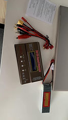


The most rewarding moment came during outdoor trials in Hanoi. Seeing the system respond to changing dust levels made the hours spent designing and debugging feel worthwhile. Our tests recorded 95% trigger accuracy, a response time of under two seconds, and a 20–25% reduction in PM2.5 concentration after each spraying cycle.


What stayed with me most was the realization that engineering could turn an invisible problem into something I could actively address. Dust pollution may be difficult to see clearly, but its impact on people's lives is real. Through this project, I learned to connect design, programming, and experimentation toward one goal: building technology that does not merely work, but works to make our surroundings healthier.
Medical Equipment Intern
Hanoi Medical University Hospital
Walking into Hanoi Medical University Hospital as a medical equipment intern, I felt both excited and humbled. I had explored respiratory diagnosis through artificial intelligence, but I wanted to understand what happened beyond the X-ray image on a computer screen: the machines that captured it, the equipment that supported breathing, and the engineering that helped keep patients safe.


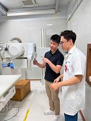


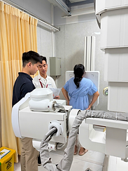

Under the supervision of three hospital engineers, I received one-on-one training in digital X-ray systems, lung ultrasound machines, invasive and non-invasive ventilators, oxygen concentrators, patient monitors, and spirometry devices. Each machine revealed a different layer of respiratory care. I began to appreciate that behind every clinical measurement was a system of sensors, electrical connections, and carefully calibrated components working together.


My most valuable lessons came from hands-on preparation. I helped assemble breathing circuits, check oxygen and air connections, inspect pneumatic and electrical systems, and prepare patient-monitoring accessories. Recording parameters such as FiO₂, PEEP, SpO₂, FEV₁, and FVC taught me that even a small technical detail could carry important clinical meaning. Under professional supervision, I also supported component-level installation and calibration checks, learning to approach each task with patience and precision.


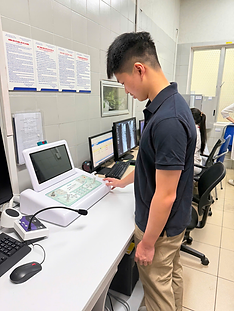


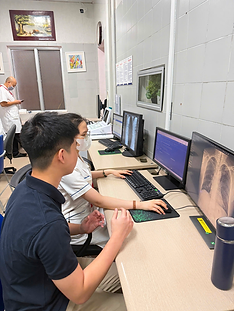

This internship changed how I understood healthcare technology. I no longer saw medical devices merely as sophisticated machines; I saw them as links between engineering and human life. I left with a deeper respect for the people who keep these systems functioning—and a stronger determination to build technology that makes respiratory care safer, more reliable, and more accessible.


